Medical Radiotherapy


Every day, thousands of people are newly diagnosed with cancer. Some of them will die within a few months, but others will be cured and live on for many more years. One of the great missions of medical science is to continue shifting this balance towards survival by learning how to treat more and more kinds of cancer successfully .

Currently, x-ray treatments (photon therapy) are very widely used, often in combination with surgery and chemotherapy. Treatments based on nuclear effects using neutron beams, proton beams, ion beams, pion beams, or neutron capture have been experimented with for a number of years, and some methods are now beginning to come into routine clinical use. In many cases, these new cancer treatment methods require advanced nuclear data and advanced schemes for calculating the transport of radiation through the body and through the components of the treatment devices themselves. Our Group is participating in these efforts.

Photon Therapy


X-rays are just high-energy photons, essentially the same as light. For medical purposes, they are normally produced by accelerating a beam of electrons to a fairly high energy (say 70 MeV) and allowing them to strike a metallic target. The x-rays produced by this interaction go out in all directions, but one beam of x-rays is selected by a collimator system and delivered to the patient. The unused x-rays are absorbed by shielding.

Most of the x-rays interact with the electron cloud of the atom and lead to ionization along the path of the beam. It is the energy deposited along the beam track by this ionization that can damage and kill the cells of the cancer. Of course, this energy can also damage or kill the normal cells of the body that lie between the x-ray source and the tumor or that lie beyond the tumor. Luckily, normal cells can take more radiation then cancer cells. The trick in constructing a photon-therapy plan is to maximize the energy deposited in the tumor while minimizing the damage to normal tissue.

A classical way to do this is to rotate the patient, just as you rotate the meat on the rotisserie of your barbecue at home. With the tumor at the center of rotation, it receives an exposure at every rotation position. But the exposure of the skin, muscle, and bone along any angle out from the tumor is only a fraction of the total dose. In more modern times, we have learned how to make smaller x-ray sources that can be rotated around the patient, but the principle is the same.

There is only one nuclear issue connected to photon therapy. A very small fraction of the high-energy photons can interact with the nucleus instead of with the electron cloud of the atom. These are called "photonuclear reactions," and they can produce neutrons. Neutrons, because they have no charge, can penetrate through matter easily. Therefore, they can produce collateral damage to healthy tissue far from the tumor site. It is important to be able to quantify this damage when constructing a treatment plan. Unfortunately, the nuclear data for photonuclear reactions have not been collected and evaluated as extensively as those for neutron reactions. Also, much more sophisticated radiation transport calculations would be required to fully evaluate the neutron dose than those methods that radiologists are currently using for photon doses. Our group is working on methods for evaluating photonuclear cross sections and preparing tables for use in radiation transport calculations. Other groups at Los Alamos, Livermore, and elsewhere, are working on improved Monte Carlo transport codes linked to CAT scans and MRI maps and tailored to the needs of the medical community.

Proton Therapy


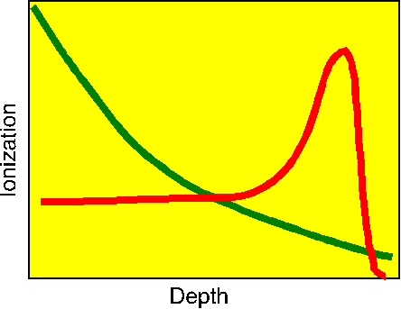
One of the problems with x-ray therapy is that it is very difficult to localize the exposure to the cancerous organ without causing dangerous exposures to surrounding healthy tissue. Ever since the early cyclotrons were built in the 40's, it has been recognized that using high-energy protons for radiotherapy could get around these problems of localization. Protons can be focussed using magnetic fields, but photons cannot. This makes it possible to generate narrow proton beams with very little spread. The shape of the beam can even be adjusted to match the shape of the target organ. Equally important is a phenomenon called the "Bragg peak." Protons slowing down in matter cause ionization along their path, but as they get slower due to this energy loss, they become even more effective at causing ionization. Eventually, they lose all their energy and stop. The result of this process is an energy deposition curve like the one shown below with a red line.

Plot of Bragg peak

Note that most of the energy deposition comes near the end of the track. This is to be contrasted to the energy deposition curve for an x-ray (the green curve), where most of the energy is deposited early in the track (in the skin and muscle). The range of the proton can be adjusted by varying the energy of the beam so that most of them deposit most of their energy in the cancerous organ. Things can even be arranged so that the energy varies through an exposure, thus scanning the energy peak across the depth extent of the tumor.

This ability to localize the damage in the cancerous organ is clearly exciting. However, to make protons penetrating enough to reach deeply into the body, they have to be made quite energetic, with perhaps as much as 250 MeV of energy. Protons with these high energies can induce nuclear reactions in addition to the simple atomic slowing down discussed above. These nuclear reactions can deposit significant amounts of additional energy at the reaction site, and they can produce secondary particles, such as neutrons. The secondary neutrons, being neutral particles, can travel far from the reaction site and induce additional reactions. Thus, we have the possibility that the nuclear effects could enhance the treatment, or that they could cause significant collateral damage.

Our Group is working to provide new sets of nuclear data for neutrons and protons interacting with biological elements for energies up to 250 MeV. Clearly, the promise of proton therapy will require very sophisticated treatment planning methods. Livermore and Los Alamos both have programs to adapt some of the methods developed over the history of the weapons program to the needs of radiotherapy professionals. The general approach is to develop methods to take diagnostic scans from CAT or MRI machines and use them to construct a three-dimensional model of the target organ and surrounding structures. This model will then be used in a Monte Carlo transport calculation that follows the atomic slowing down and the nuclear reactions for all particles in full detail. Sophisticated graphical displays of the results would then become available to the radiologist in real time, thus allowing the exposure to be optimized with the patient in place. This work pushes the boundaries of nuclear data, radiation transport, computer technology, and interactive interface design. But it promises great payoffs in saved lives.

There are several sites on the Web with additional information on proton therapy and discussions of the work at particular medical centers.

Other Particle-Therapy Options


Radiation therapy using protons is discussed above. It is also possible to substitute heavier ions, such as helium nuclei (alpha particles) for the protons. Even heavier ions, such as oxygen or nitrogen could be used, and that would open up the possibility of chemical effects from the stopped ions. The use of heavy ions for cancer therapy would also require the development of additional nuclear data libraries.

Boron Neutron-Capture Therapy


The Holy Grail of medicine is the "magic bullet;" a treatment that goes directly to the site of a problem, fixes it, and then politely disappears. Some people think that "Boron Neutron-Capture Therapy" (BNCT) could be a magic bullet. When 10B captures a low-energy neutron, it goes through the reaction

n + 10B → 4He + 7Li + 2.7895 MeV

The ranges of the energetic alpha particle and lithium nucleus are very short; typically the size of a cell. Besides, the neutron absorption cross section for B-10 is very large--much larger than most of the nuclei normally found in the cell. Therefore, if you could get some B-10 into a cancerous cell, and then expose it to a flux of neutrons, you could kill the cell with a minimum of collateral damage.

To verify the energy release of this reaction for yourself, jump to the qtool, enter 1 for the projectile (neutron), 5010 for the target (B-10), and 0 for EMAX.

There are several areas of active research related to BNCT. One is to find boron compounds that will concentrate in cancerous organs. Maybe a monoclonal antibody that only goes to a cancerous prostate? The second is to find hospital-scale methods to provide beams of epithermal neutrons with enough energy to penetrate to the target organ and with little fast-neutron contamination. These could be special fission reactors, or perhaps accelerator-based systems. There are few needs here for advanced nuclear data, but we watch with interest.


Start the schoolbus tour
ryxm@lanl.gov
14 March 1997 (updated May 2012)